How Dermatologists Treat a Microblading Granuloma: Biopsy, Steroid Injections, and Realistic Timelines
Published on August 22, 2026
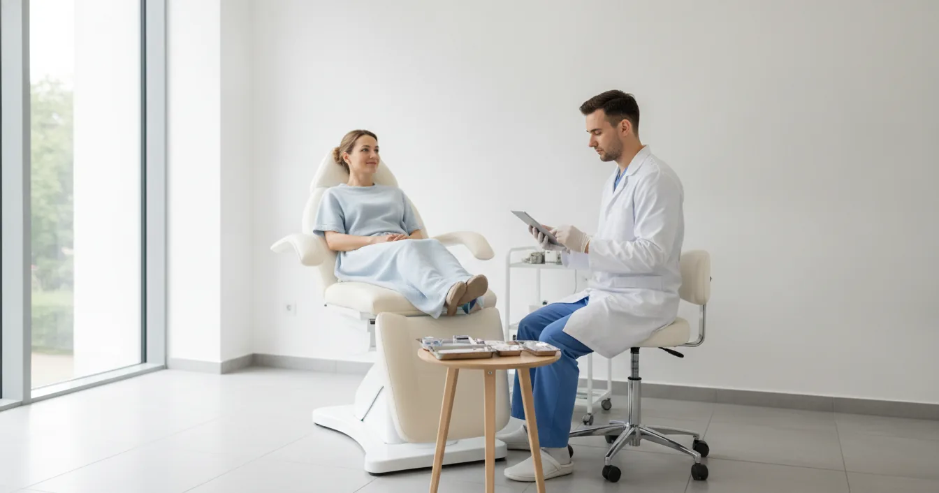
If you have already worked out that the firm lumps in your brows are a delayed pigment reaction rather than a healing spot, the next question is a practical one. What does treating it actually involve? Our guide to bumps, lumps, and granulomas after microblading covers how to recognise one and why it showed up months or years after the appointment. This is the other half: the appointments, the needles, the prescriptions, the bills, and how long each stage realistically takes. None of it is dramatic, but little of it is fast. Knowing the shape of the process in advance makes the waiting much easier to manage.
Book a Dermatologist, Not a Beauty Clinic
The single most common wrong turn is going back to the artist. A permanent makeup artist cannot diagnose a granuloma, cannot prescribe, and cannot inject, and in most jurisdictions it would be well outside their licence to try. The same goes for a medspa offering to laser the pigment out. What you need is a board-certified dermatologist, ideally one who has seen tattoo reactions before, since brow pigment reactions are uncommon enough that general practice tends to miss them.
Bring three things to the appointment. First, dated photographs showing when the bumps appeared and how they have changed. Second, whatever paperwork you have from the procedure, including the pigment brand, colour name, and batch number if your artist recorded them, which good studios do. Third, a short written timeline of your own symptoms. Lead with the microblading history the moment you sit down, however long ago it was, because that one detail is what separates a correct diagnosis from a course of acne treatment that will not work.
The First Visit and the Punch Biopsy
Expect the first visit to be mostly examination and history. The dermatologist will look at the lesions closely, often with a dermatoscope, feel their firmness and depth, and ask about pigment colours, other tattoos, previous reactions, medications, and any systemic symptoms. A firm red-brown papule sitting exactly along the pigmented strokes is suggestive, but suggestive is not the same as diagnosed. Foreign-body granulomas, sarcoid-type granulomas, lichenoid reactions, allergic responses, and atypical infections can all look similar to the eye, and they do not respond to the same treatment.
That is why a punch biopsy is usually offered. A small circular blade, typically three or four millimetres across, removes a full-thickness core of skin from one representative lesion after the area is numbed with injected local anaesthetic. The numbing sting is the worst part. The punch itself takes seconds, and the site is closed with a single stitch or left to heal on its own, with results back from pathology in roughly one to two weeks. Under the microscope the pathologist can see whether the immune cells are organised into non-necrotizing granulomas around pigment particles, which is precisely what a published case of eyebrow bumps three months after microblading showed.
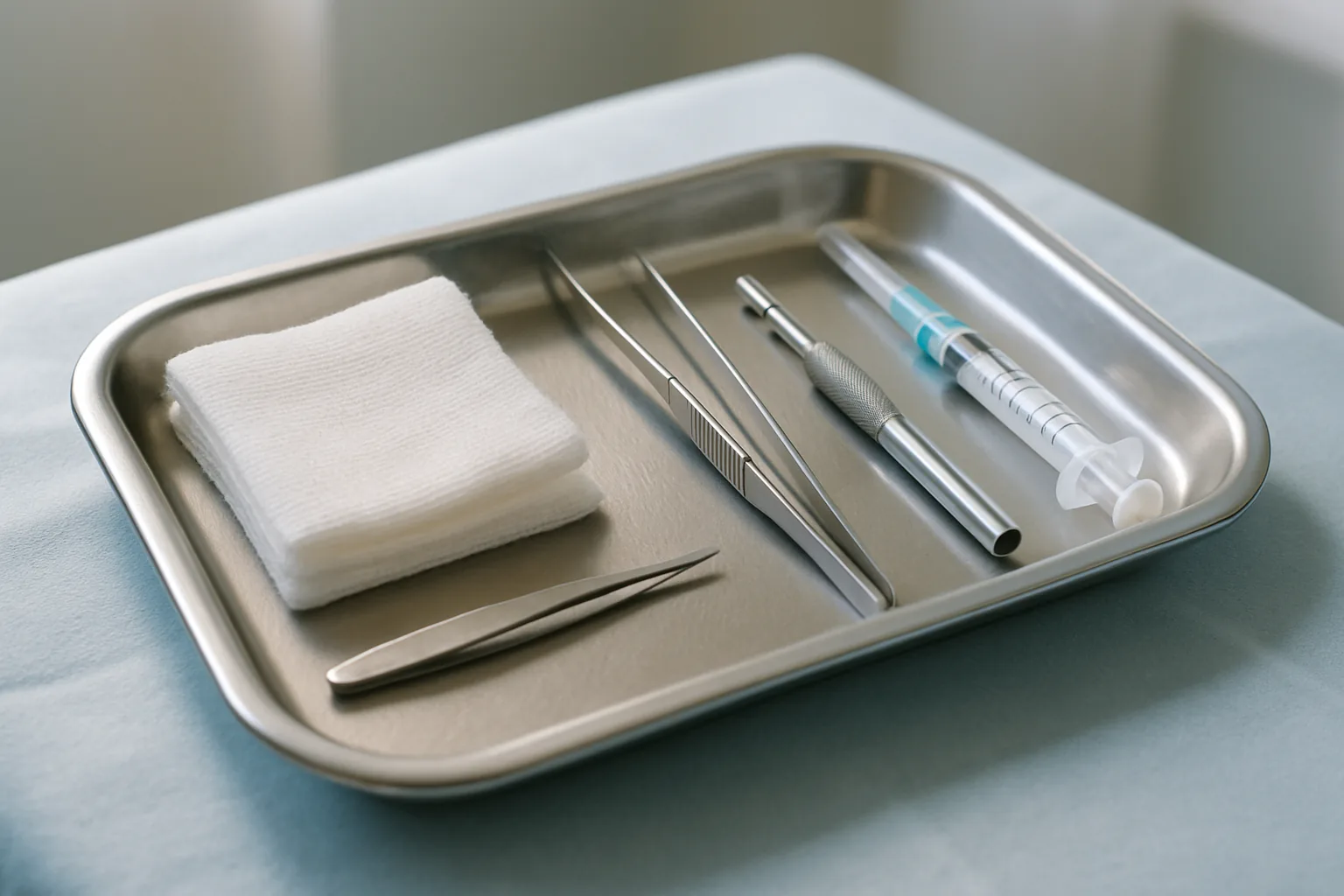
There is a real trade-off worth raising out loud. A biopsy on the brow leaves a small permanent mark and a gap in the pigment, and it may disturb brow hair at that spot. Ask the dermatologist to take the sample from the least visible lesion, usually toward the tail of the brow. Most people conclude the certainty is worth it, because the treatment path genuinely diverges depending on what comes back.
Screening Beyond the Skin
If the biopsy shows granulomas, expect the workup to widen. Chronic inflammatory reactions to black pigment carry a meaningful rate of involvement elsewhere in the body. In an analysis of 308 tattoo and permanent makeup complications, extracutaneous involvement was found in 21.4 percent of chronic inflammatory black tattoo reactions, including tattoo-associated uveitis in 7.1 percent and systemic sarcoidosis in 14.2 percent. That is why a chest X-ray, bloodwork, and often a referral for an eye exam get added. Brow pigment is overwhelmingly black, so this applies to a great many microblading cases.
Most of these come back clear, as they did in the case above, where blood tests and chest imaging were normal. But the screening is not defensive box-ticking. Sarcoidosis found early through a brow lump is a genuinely useful diagnosis, and our guide to microblading with an autoimmune condition explains why that association runs in both directions.
Intralesional Steroid Injections: How the Rounds Work
Corticosteroids are the workhorse of treatment, and for firm, established lumps the delivery method that matters is intralesional injection: steroid, usually triamcinolone acetonide, placed directly into the lesion with a very fine needle. Injecting into the lump means a high local concentration where the inflammation is, without the systemic exposure of tablets.
Three things are worth knowing before the first round. It is done at a dilute concentration on the face, often in the range of 2.5 to 5 mg/mL rather than the stronger mixes used on the body, because facial skin is thin and over-treatment causes visible problems. It is uncomfortable in a pressured, stinging way for a few seconds per injection point, and the whole appointment takes minutes. And it is almost never a single visit. Sessions are typically spaced four to six weeks apart, and two to four rounds is a common course, with the dermatologist reassessing flattening at each visit before deciding whether to repeat. The tattoo-reaction literature relies on steroids because they can be repeated and reassessed, and that is the realistic picture here: repetition, not a single fix.
The side effects to watch for are local. Too much steroid in one place can thin the skin, leaving a dented or shiny patch, and it can lighten the skin around the injection site, which is more noticeable and slower to recover on deeper skin tones. Both are reasons a careful dermatologist under-treats on the first round and builds up.
Topical and Oral Options
Milder or flatter reactions often start with a potent topical steroid such as clobetasol, applied in short bursts rather than continuously, since the same thinning risk applies to skin around the eye. A topical calcineurin inhibitor is sometimes used as a steroid-sparing alternative for longer stretches. If itch is a major complaint, particularly in allergic-type reactions, oral antihistamines help with the symptom while the underlying treatment works.
When granulomas resist injections, dermatologists move to systemic drugs borrowed from sarcoidosis and other granulomatous disease: hydroxychloroquine, tetracycline-class antibiotics used for their anti-inflammatory effect rather than as antibiotics, allopurinol, and in stubborn cases methotrexate. These are prescription medicines with monitoring requirements, including eye checks on hydroxychloroquine and blood tests on methotrexate, and they take months rather than weeks to show effect. Tell your dermatologist if you are pregnant, nursing, or trying to conceive, because methotrexate is absolutely contraindicated in pregnancy and it narrows the list sharply. Pregnancy narrows the options on the artist’s side of the chair too, for related reasons set out in our guide to microblading while pregnant or breastfeeding.
Realistic Timelines
Set your expectations in months. Some reactions resolve with no treatment at all: in the microblading case documented in the Dutch literature, the papules disappeared spontaneously within six months. If your reaction is asymptomatic and not spreading, watchful waiting for a few months is a legitimate medical choice rather than a brush-off.

With treatment, a rough map looks like this. Diagnosis and biopsy results take two to four weeks from the first call. A steroid injection course runs three to six months across its two to four rounds. Systemic medication is judged over three to six months before anyone calls it a failure. And the pigment itself does not leave, so a granuloma that flattens completely can still recur later, especially after sun exposure, illness, or any attempt to break the pigment down. The honest framing is that 91.9 percent of tattoo complications in that 308-case series were chronic rather than acute. You are managing a long-running reaction, not curing an infection.
What It Costs, and What Insurance Covers
Prices vary widely by country, region, and clinic, so treat these as general guidance rather than quotes. In the United States, a specialist consultation without insurance commonly runs somewhere around 150 to 350 dollars, a punch biopsy with pathology roughly 200 to 500 dollars, and an intralesional steroid session roughly 75 to 250 dollars per visit, which multiplies across a course.
The insurance question has a better answer than most people expect. A biopsy-confirmed granuloma is a medical diagnosis, not a cosmetic complaint, and diagnosis and treatment of a complication are usually billable as medical care even though the microblading that caused it was cosmetic and certainly is not. Ask the clinic to confirm the coding before treatment. What insurers reliably will not cover is anything framed as improving the appearance of the brows, including pigment removal, which is worth knowing before you plan around it. Our two-year cost breakdown covers the budgeting side of the procedure itself.
Why Laser Removal Is the Last Resort
It seems logical: the pigment causes the reaction, so remove the pigment. In practice, lasering a reactive tattoo can make things sharply worse. Fragmenting the pigment releases more of it into contact with the immune system, and there are documented cases of an allergic flare appearing partway through removal, once the laser reached a colour that had been sitting quietly underneath. Laser removal also carries its own complication list, including blistering, scarring, and dyspigmentation, and those risks rise steeply with an untrained operator. On already inflamed brow skin, scarring risk is not theoretical.
None of this makes removal impossible. It means removal is a decision made with a dermatologist after inflammation is controlled, sometimes under steroid cover, with a test patch first, and never as the opening move. Our guide to removing bad microblading walks through how laser and saline compare when pigment removal is the goal.
Between Appointments
There are a few genuinely useful things to do while you wait. Photograph the brows in the same light every couple of weeks so change is measurable rather than remembered. Use sunscreen on the area, since ultraviolet exposure can aggravate pigment reactions. Leave the lumps alone, because squeezing a granuloma achieves nothing except inflammation. Keep skincare bland: no acids, no retinoids, no scrubs on the brow while it is reactive. And do not get any further pigment work anywhere, including a touch-up on the same brows, until the reaction has been diagnosed and settled.
The reassuring part is that these reactions are uncommon, most stay confined to the skin, and a substantial number either resolve on their own or respond to steroid. The frustrating part is the pace. Bringing a clear history to a dermatologist early, accepting the biopsy, and viewing treatment in months rather than weeks can make the process more manageable.
Sources
- Journal of Cosmetic Dermatology on what 308 tattoo and permanent makeup complications revealed about chronic reactions
- Nederlands Tijdschrift voor Geneeskunde for the brow biopsy that found sarcoid granulomas after microblading
- Missouri Medicine with the management strategies used across tattoo-related skin reactions
- Current Problems in Dermatology covering the late-onset complications specific to cosmetic tattooing
- Dermatologic Surgery documenting a delayed granulomatous reaction traced to eyebrow microblading
- Journal of Clinical Medicine on an allergic flare provoked partway through eyebrow pigment removal
- Dermatology for the complications practitioners report during tattoo removal