Microblading Over a Mole or Near a Skin Lesion: Why Artists Work Around Them and When to See a Dermatologist First
Published on September 27, 2026
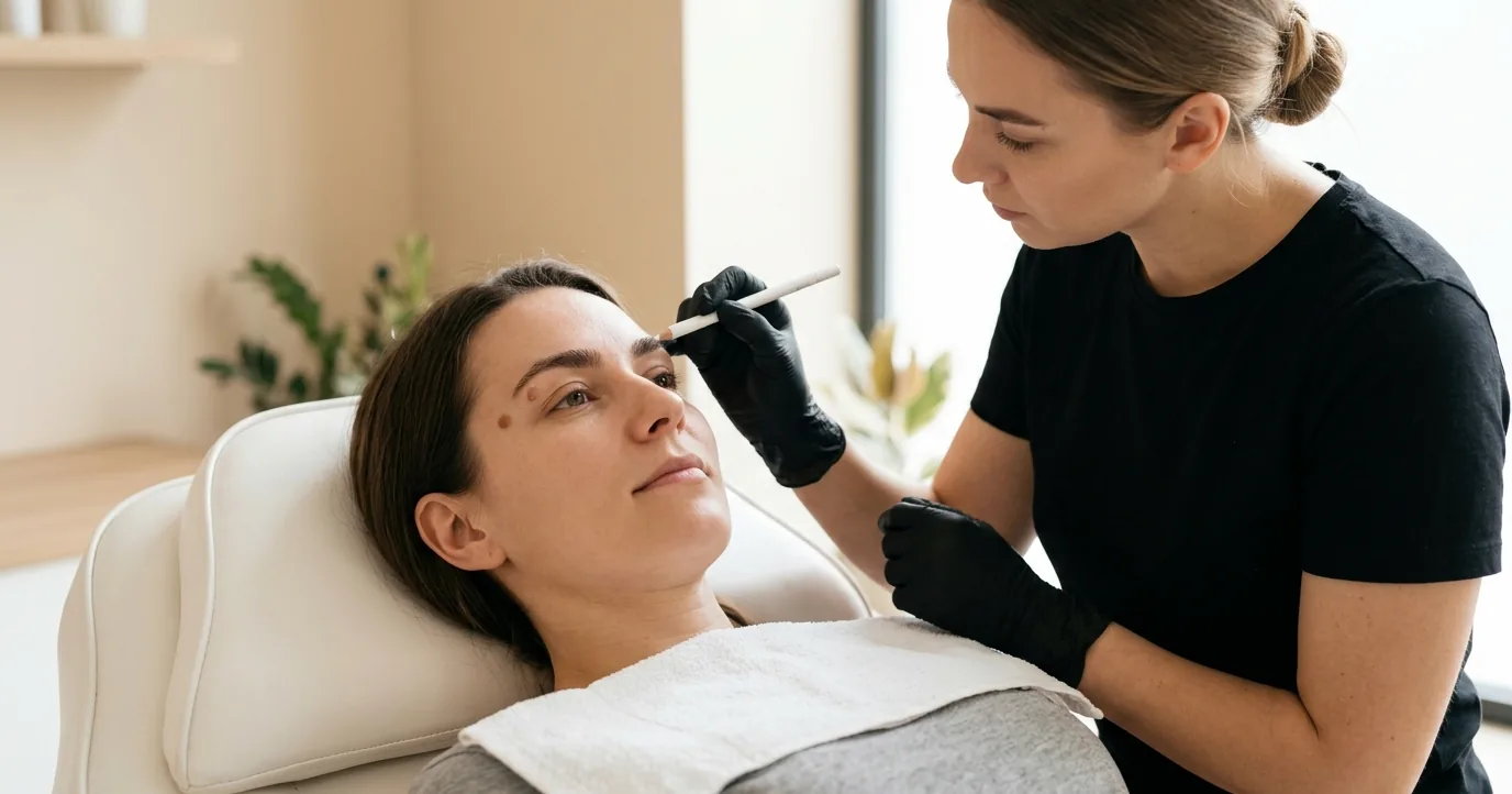
It happens at consultations more often than clients expect. The brow hairs get brushed up, the lamp comes down, and the artist stops at a small brown spot in the arch, or at a pearly bump near the tail that the client has half noticed for a year. The next question is nearly always the same: can’t you just go over it?
No. A careful artist will work around it or send you to have it checked first, and the reason has little to do with the spot being in the way. It is about what pigment does to everyone’s ability to read that spot later, which is also why some spots need a dermatologist before they need a brow artist.
What Actually Turns Up in the Brow Line
Most of what an artist finds is harmless. Ordinary moles, seborrheic keratoses (the waxy, stuck-on growths that become common with age) and skin tags all turn up around the brows and eyes, and on and around the eyelids the American Academy of Dermatology (AAD) notes that most growths are benign.
Most, not all. Actinic keratoses, rough scaly patches of sun damage that can progress to cancer, occur most often on the face. Basal cell and squamous cell carcinoma can appear as a sore that does not heal, or as skin that is shiny and pearly, firm and scar-like, or scaly and crusty. Melanoma often first shows as a change in the shape, colour, size or feel of an existing mole, which is what the ABCDEs track: asymmetry, an irregular border, uneven colour, a growing diameter, and evolution over weeks or months. The eye area also hosts a rarer one. Sebaceous carcinoma is aggressive, most often starts on an eyelid, where oil glands are most densely packed, and can appear as a painless, round, firm lump.
The brow itself is not exempt. Published case reports describe basal cell carcinoma in the brow region of five women aged 21 to 43, so this is not only an older client’s problem. They describe a squamous cell carcinoma on an 81-year-old woman’s eyebrow that was treated for three months as a wound slow to heal because of her diabetes, and a sebaceous carcinoma that started in a 68-year-old man’s eyebrow, grew over ten months, and proved fatal after he declined the wider surgery his doctors recommended.
Brow hair makes all of this easier to miss. A spot under hair can sit unexamined for years, and a consultation with the hairs brushed back under a bright lamp may be the closest look that skin has had in a long time. Nobody can tell a harmless spot from a dangerous one by eye in that moment, a skilled artist included. That call belongs to a dermatologist, and sometimes to a biopsy.

Why Pigment Should Never Go Over a Mole
Brow pigment is not the suspected cause of these cancers. A 2025 Italian case series and review of 45 melanomas found on tattooed skin judged the association coincidental rather than causal, and the AAD says more research is needed to know whether tattoos and skin cancer are linked at all. (What pigment does once it is inside you is a separate question, covered in our look at where brow pigment goes and what the cancer research shows.) The concern dermatologists raise is more practical. In the AAD’s words, “the greater risk is that a tattoo may disguise skin cancer and make it harder to detect.” That happens in three ways.
Ink changes what the spot looks like. Border and colour are two of the ABCDEs, and both can change once a spot has been tattooed. A 2024 study from Porto Alegre, Brazil, examined 112 tattooed people and found 194 moles sitting under ink. Seen through a dermatoscope, the lighted magnifier used in skin checks, the pigment blurred the mole’s structure in more than half of them. In almost two-thirds it partly or completely obliterated the mole’s own pattern, which can hide exactly the asymmetry a skin check looks for, and in about one mole in six it produced a blue veil, a pattern dermatologists associate with melanoma.
Ink can make a harmless mole look dangerous. The same team notes that a mole traumatised by tattooing can develop cell changes that raise suspicion under the microscope, so ink in a mole can either delay a real diagnosis or prompt surgery that was never needed.
Ink gets in the pathologist’s way. If a tattooed spot is biopsied, the pigment can make cancer cells harder to pick out in the sample. Each of these costs time, and time is what makes a skin cancer more serious by the point it is caught.
The specialist literature is blunt about the conclusion. A Duke University review of 160 tumours arising in tattoos said tattoos should never be placed over existing moles or precancerous lesions. German laser specialists, reporting a melanoma that developed in a mole inside a tattoo, wrote that tattoos should never go on pigmented lesions at all.
None of that research was done on brows, but it transfers directly. The FDA counts permanent makeup, eyebrows included, as a type of tattoo, and many brow pigments lean on dark browns and black, one of the three ink colours the AAD singles out as most likely to disguise a worrying spot. Microblading fades, yet it stays in the skin for years, which is a long time for a spot to change under cover. One Korean case shows what that looks like: a 48-year-old woman whose brows had been tattooed six years earlier noticed a black mass in her right brow and waited a year before having it seen. It was a basal cell carcinoma, removed in two surgical stages. In a tattooed brow, a dark bump is easy to dismiss as a little extra pigment.
What a Careful Artist Does at the Consultation
This is where a licensed artist earns the fee, and it is one more reason unlicensed practitioners are a genuine safety risk: often nobody is looking at the skin at all. Good practice looks like this.
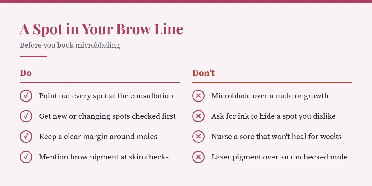
- Look at the bare skin. Brush the hairs up, use good light, and record every mole, bump, scaly patch or sore on the intake form, with a photo if the client agrees.
- Work around, never over. Pigment stays off moles, keratoses, skin tags and anything undiagnosed. For body tattoos, dermatologists recommend 0.5 to 1 cm of clear skin around a mole. That is a large share of a brow’s height, so a mole in the body of the brow usually means reshaping the design, leaving a gap that natural hair can cover, or a dermatology appointment first.
- Refer, don’t reassure. “That looks like a normal mole” is a diagnosis. The honest line is “I can’t tell what that is, and I won’t work near it until a dermatologist has seen it.”
- Never camouflage. Clients sometimes ask for pigment over a spot they dislike. That is the one request to refuse outright, because hiding the spot is precisely the harm.
- Leave removal to doctors. Snipping off a skin tag or shaving a mole is a medical procedure, done in a clinic, with the tissue usually sent to a lab.
A patch test still belongs in every consultation, but it tells you how your skin reacts to the pigment and nothing about a mole. Spots on the brow sit alongside the other reasons to delay in our complete guide to microblading risks and contraindications.
The Simple Rule: A Dermatologist Before the Booking
See a dermatologist before the appointment, not after, about any spot in or near the brow that is:
- new, or new to you
- changing in shape, colour, size or feel
- itching, bleeding, oozing or crusting
- a sore that has not healed, or heals and comes back
- a lump near the eyelid that stays despite treatment
Book a full skin check first even without a specific worry if you have had skin cancer before, have a family history of it, have many or unusual moles, or have a weakened immune system. The AAD and the Duke reviewers both single out these groups for a dermatologist’s assessment before any tattoo.
The order matters. Before the appointment, the dermatologist sees the spot as it really is. Afterwards, they see it through pigment that will sit there for years, and any biopsy gets cut through brows you have just paid for. That creates its own hesitation: in the Italian melanoma series, a man with a suspicious mole under his tattoo hesitated over removal because the excision would damage a design he cared about. And don’t nurse a brow sore yourself for weeks; the squamous cell carcinoma above spent three months being treated as a slow wound.
Microblading After a Mole or Lesion Is Removed
If a spot should come off, how long you wait depends on how it was removed and what the lab finds. Raised growths are often shaved or snipped off, and keratoses frozen with liquid nitrogen. When cancer is a concern, a full-thickness excision takes a margin of normal skin, about 3 to 4 mm or more, and is closed with stitches; certain skin cancers get Mohs surgery, which spares as much healthy skin as possible. Removed tissue usually goes to a lab.
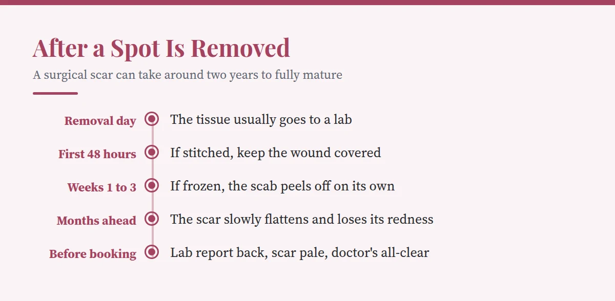
First, the lab report. Book nothing until it is back. If the spot was benign, the only remaining question is healing. If it was a skin cancer, your dermatologist or surgeon sets the timeline, and the site may need to stay visible for follow-up. The AAD advises keeping ink off any area your dermatologist wants to check at future appointments, and the Italian team behind the melanoma review discourages tattoos over melanoma surgical scars in particular.
Then, surface healing. A stitched wound is usually kept covered for the first 24 to 48 hours, and the stitches come out at a follow-up visit. After freezing, a blister often forms within hours and the scab peels away on its own in one to three weeks; leave it alone. If the spot does not look gone once everything has healed, that is a call to the surgeon, not the artist.
Then, the scar. A closed wound is not a settled one. An NHS hospital’s scar care guide says a surgical scar can take around two years to mature fully, flattening and losing its redness gradually, and warns that scar tissue has less protection from the sun, so shield it with sunscreen meanwhile. Scar tissue also takes pigment less predictably than healthy skin, often healing patchy or blurred, as our guide to microblading for medical brow loss explains. Get the site cleared by the doctor who removed it, and expect a careful artist to wait until the scar is flat and pale rather than pink.

If a Mole Is Already Under Your Brow Pigment
Plenty of people had their brows done before anyone raised this, and there is no reason for alarm. Most moles never become anything, and melanomas in tattooed skin are rare enough that the medical literature counts them in dozens. What changes is how you keep watch.
Tell your dermatologist the area is tattooed, include your brows in regular skin checks, and take dated photos in the same light every few months so change is easier to see. The AAD suggests paying extra attention to skin tattooed in black, blue or red ink. A new lump in the strokes months or years later can be a granuloma, a reaction to the pigment itself, which our guide to bumps, lumps and granulomas after microblading covers. It can also be something else, and only a dermatologist can tell the two apart.
Laser removal needs particular care. Clearing the pigment can uncover a mole that has been hidden for years, and the German melanoma case developed while the tattoo over it was being lasered off. Those authors advise excising a pigmented lesion before laser removal starts, and the AAD recommends having laser removal done by a dermatologist, who can check any spot the laser reveals even if a biopsy means pausing sessions. If you are weighing lasers, our comparison of picosecond, Q-switched and ruby lasers for brow removal covers the rest.
The Bottom Line
A spot in your brow line is not a reason to give up on microblading. It is a reason to change the order: dermatologist first, artist second. Artists work around moles because skin kept clear of pigment stays readable, for you and for every dermatologist who looks at it later. Results still depend on the artist’s skill and your skin type, and microblading is semi-permanent rather than permanent, but pigment over a spot can leave it unwatched for years. No brow shape is worth that trade.
Sources
- American Academy of Dermatology on how a tattoo can disguise skin cancer, and why to work around moles
- AAD for the sebaceous carcinoma overview and why it favours the eyelids
- National Cancer Institute with the ABCDE rule and the mole changes worth reporting
- NCI on the signs of basal cell carcinoma, squamous cell carcinoma and actinic keratosis
- Watanabe, Dantas and Bonamigo on what tattoo pigment does to the dermoscopic picture of a mole
- Lebhar and colleagues for the Duke review of 160 tumours found in tattooed skin
- Brusasco and colleagues with 45 melanomas on tattooed skin and the case against a causal link
- Pohl, Kaiser and Raulin on a melanoma that developed in a tattooed mole during laser removal
- Han, Jo and Kim describing a basal cell carcinoma in a brow tattooed six years earlier
- Dzubow for basal cell carcinoma in the brow region of five younger women
- Hamamin and colleagues on an eyebrow squamous cell carcinoma first treated as a slow wound
- Ye and Li with a fatal sebaceous carcinoma that began in the eyebrow
- MedlinePlus on the ways a skin lesion is removed and sent for testing
- MedlinePlus for caring for stitches, open wounds and frozen spots after removal
- Royal National Orthopaedic Hospital on how long a surgical scar takes to mature
- FDA for permanent makeup, eyebrows included, as a type of tattoo